Shopping Cart


")


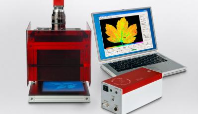
, obtained with a plant of Arabidopsis thaliana in actinic light")

")


")
")
")
")


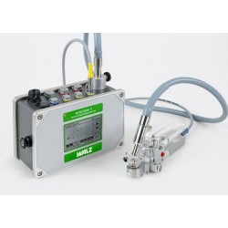
Chlorophyll Fluorescence System WALZ
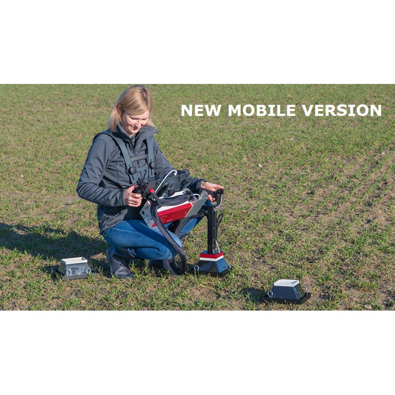
MOBILE Version is dedicated to chlorophyll fluorescence imaging in the field.
The MAXI and MINI versions may be combined with the Gas-Exchange System GFS-3000 thus integrating detailed spatial information on photosynthesis and with exact analysis of photosynthetic carbon fixation.
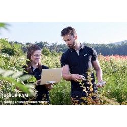
OUTDOOR Version:
For field research on leaves, small plants, ground vegetation, and photosynthetic crusts (2.4 x 3.2 cm)
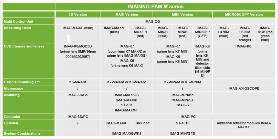
The IMAGING-PAM family comprises the versions MAXI, MINI and MICROSCOPY. The different versions employ the same Multi Control Unit IMAG-CG. Also, the same camera can be used for different versions. This modularity permits easy and most cost-effective switching between various applications and magnifications provided by the IMAGING-PAM M-Series.
The MAXI and MINI versions can be combined with the Gas-Exchange System GFS-3000 and, thus, integrates detailed spatial information on photosynthesis with exact analysis of photosynthetic carbon fixation.
Most recently, an advanced method to create three-dimensional plant images has been added to the MAXI version. This new configuration opens the way to project data of fluorescence analysis on the three-dimensional plant image so that photosynthesis can be analyzed in the context of whole plant architecture.
Walz provides a family of chlorophyll fluorescence imaging systems, the IMAGING-PAM M-Series for imaging large scale samples with areas exceeding multiwell plate format as well as microscopically small samples at the level of single cells or even chloroplasts.
The M-Series instruments have been used with remarkable success in such diverse fields of science as plant phenotyping, leaf ecophysiology, coral research, phytopathology and marine ecophysiology.
All members of the IMAGING-PAM M-Series family: MAXI, MINI and MICROSCOPY version can share many components like cameras and the Multi Control Unit IMAG-CG. That makes it easy and most cost effective to switch between different applications and magnifications of the IMAGING-PAM M-Series.
New: High speed GigE Vision camera connection.
Combination of MAXI and MINI versions with Gas-Exchange System GFS-3000 extend the analysis options.
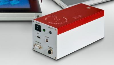
Design: Aluminum housing featuring built-in Li-ion battery, sockets for cable connections with CCD Cameras IMAG-K6 or IMAG-K7, connectors for the MAXI, MINI and MICROSCOPY Measuring Heads and Battery Charger 2120-N
Microcontroller: RISC processor
User interface: For Windows 10 PC with ImagingWinGigE Software; connection via GigE ethernet; keyboard operation; monitor screen display
Power supply: Internal rechargeable Li-ion battery 14.8 V/5.2 Ah
Power consumption: 9 W (500 mA) drawn from internal Li-ion battery
Recharging time: Approx. 4 hours (IMAGING-PAM turned off) via Battery Charger 2120-N
Operating temperature: -5 to +45 °C
Dimensions: 25 cm x 10.5 cm x 11 cm (L x W x H)
Weight: 2.1 kg (incl. internal battery)
Input: 90 to 264 V AC, 47 to 63 Hz
Output: 19 V DC, 3.7 A
Operating temperature: 0 to 40 °C
Dimensions: 15 cm x 6 cm x 3 cm (L x W x H)
Weight: 300 g
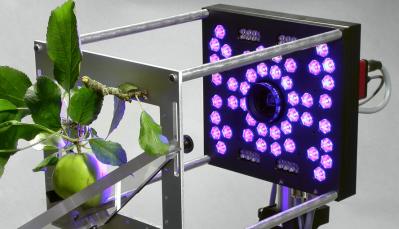
Design: LED-Array mounted on printed-circuit board in aluminum housing with central opening for CCD Cameras IMAG-K6 or IMAG-K7. The necessary cooling is realized by a fan arranged on the top side. Cable connections to the IMAG-CG control unit and the external 300 W power supply are led out of the rear of the housing
Light sources for fluorescence excitation and actinic illumination: 44 royal-blue 3 W Cree LEDs (450 nm), equipped with individual collimating optics; standard excitation intensity 0.5 μmol m-2 s-1 PAR, modulation frequency 1-8 Hz; maximum actinic intensity 1900 μmol m-2 s-1 PAR; maximum saturation pulse intensity 4000 μmol quanta m-2 s-1 PAR
Light sources for assessment of absorbed PAR and live video option:
16 red LEDs (660 nm); 16 NIR LEDs (780 nm)
Light field properties: Vertical incidence on sample; LED distribution optimized for uniformity; at standard working distance maximal deviation from mean intensity +/- 7 %
Dimensions: 18.5 cm x 18.5 cm x 4.5 cm (L x W x H)
Weight: 1.3 kg (incl. cable 1.5 m long)
Input: 90 to 264 V AC, 50/60 Hz
Output: 43 to 57 V, 5.2 A (adjusted to a voltage depending on the LED array)
Operating temperature: 0 to 40 °C
Dimensions: 22.6 cm x 11 cm x 5.8 cm (L x W x H)
Weight: 1.75 kg
Design: mechanically modified black and white C-mount camera operated in 10-bit-mode at 16 frames/sec featuring 2 x 2 pixel binning. The camera was modified for the required automatic filter slider
CCD Chip size: 2/3" (1392 x 1040 pixel primary resolution)
Interface: GigE-Vision®
Dimensions: 8,64 cm x 7 cm x 2,9 cm (L x W x H) (without objective lens)
Weight: < 200g
Design: Aluminum frame with LED projector, LED array IMAG-MAX/L, camera IMAG-K6/MOD3D, sliding door and darkening blinds mounted. Compact design
Sample position: potted plants (max. diameter 10 cm, height 4-5 cm are held in position vertically on a software-controlled turntable. For the measurement they are rotated four times by 90°, the camera viewing angle is always 0° - vertically from above.
Dimensions: 41.5 cm x 24 cm x 50 cm (L x W x H without cables)
Weight: 11.32 kg (Including stand, projector, camera, turntable, without cables)
Design: Plastic housing with LED light source (WXGA native resolution and 700 lm luminous intensity. Permanently mounted on IMAG-3D/GS
Focus: manual
Projection: DLP
Power Consumption: 75W
Weight: 510 g
Dimensions: 19 cm x 12 cm x 19 cm (L x W x H)
Operating temperature: 5 °C - 40 °C
Design: Aluminium body as carrier for 2 different calibration patterns (calibration and fusion). Three different angle positions (needed during calibration process) can be as fixed steps.
Dimension: 14 cm x 14 cm x 10 cm (width x depth x height)
Weight: 650 g (calibration body without calibration plates)
Minimum PC requirements: Windows 10 OS, Intel Core i5-9xx or higher CPU, NVDIA® graphics board (min 2 GB or more dedicated memory), SSD, 8GB RAM, internal Gigabit Ethernet (GigE), HDMI interface.
The 3D part of the software can only be used under License. Therefor a USB dongle is necessary (included in the basic instrument).
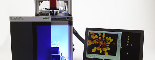
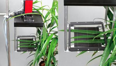
The basis of the IMAG-3D is the successful MAXI IMAGING head expanded by a 3D scanner. Using algorithms developed by the Fraunhofer Institute for Integrated Circuits IIS, the system creates a 3D mesh from images of structured light casted on the plant by a projector. The projector is mounted on the backside of the instrument whereas the camera is positioned above the plant. For 3D imaging, this camera is used in the high resolution mode (1280 x 960 pixels) to precisely digitize the sample surface. For fluorescence measurements, highest sensitivity is achieved by switching the camera to 4 pixel binning which results in larger pixel size.
A dedicated tabletop housing offers darkening for 3D and PSII analysis. The housing is optimized for high precision and cost efficiency. A software-controlled turntable enables researchers to measure single potted plants with a rosette of up to 100 mm maximum diameter. The 3D version requires only a small footprint so that it fits on almost every laboratory desk. For 3D recalibration purposes, the instrument is shipped together with two calibration patterns and a holder so that a quick calibration of the whole system is possible at any time.
Multi Control Unit
• IMAG-CG Multi Control Unit to connect Measuring Head and GigE –Vision® CCD camera as M-Series IMAGING-PAM MAXI, MINI and MICROSCOPY version including ImagingWin GigE Software.
Measuring Head
• IMAG-MAX/L (blue measuring light version, 450 nm, standard applications)
Camera and Lens
• IMAG-K6/MOD3D with increased NIR sensitivity (2/3" chip, primary resolution 1280 x 960 pixels ) with prime lens objective (F1.4/f=16 mm, c-mount). A software-controlled filter slider with ND filter for 3D and PSII fluorescence detection filter.
Mounting
• IMAG-3D/GS Stand. Darkening and rack for IMAGING-PAM 3D version
Computer
• IMAG-3D/PC is a branded notebook PC (with Intel Core i5-8xx or higher CPU, NVDIA® graphics board (2 GB or more dedicated memory), SSD, 8GB RAM, internal Gigabit Ethernet (GigE), HDMI interface, Windows 10 OS)
System Combination
• Since the instrument is designed as a compact stand-alone unit there are currently no combinations with other Walz Instruments possible except the ULM-500 universal light meter for PAR calibration
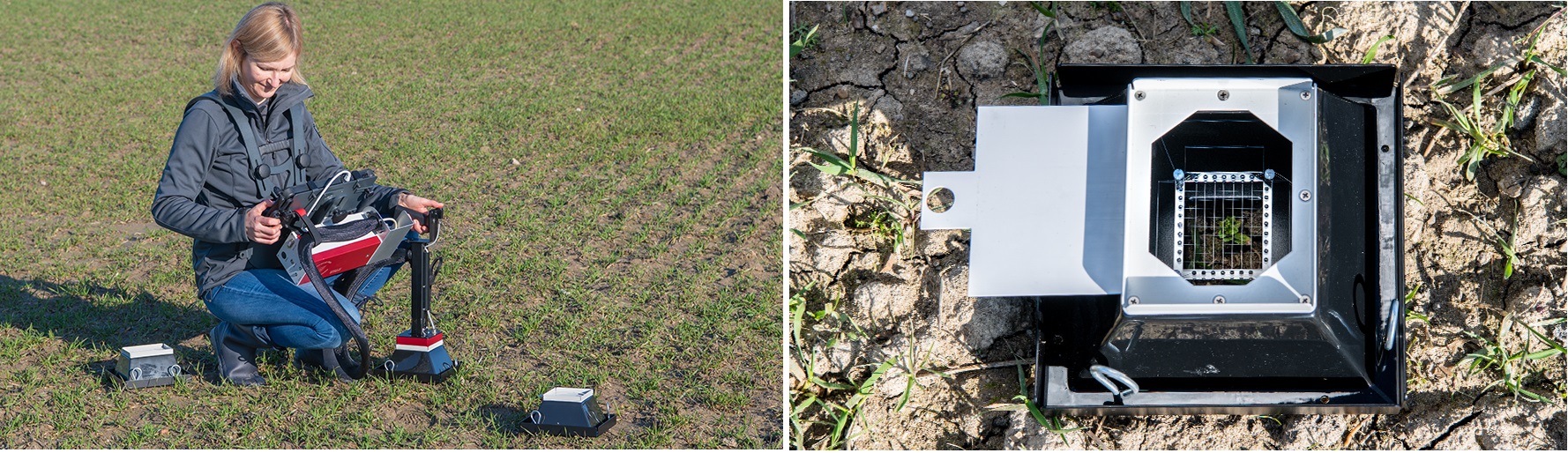
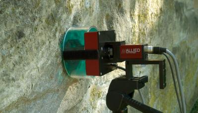
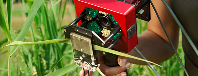
Specially developed for chlorophyll fluorescence imaging in the field. The measuring system shares features with the MINI Version of the IMAGING-PAM like the moderate power consumption and the imaging area of 24 x 32 mm.
The MOBILE version is particularly dedicated to the study of small plants, ground vegetation, and photosynthetic crusts.
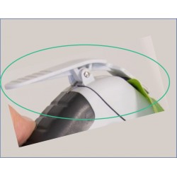
The newly developed dark acclimation box allows field measurements of the maximum photosynthetic quantum yield of photosystem II (FV / FM).
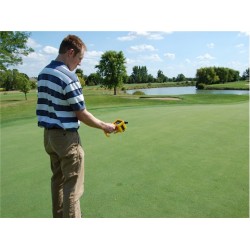
The extension handle and trigger button on the measuring head provides the system with excellent ergonomic properties.
A high level of mobility in the field is achieved by mounting the unit, the system’s sunlight-readable tablet computer and its control unit on a belly carrying tray. The carrying system may easily be adapted to the user's individual physical dimensions and height.
As an example of application, please see the journal "Precision Agriculture". The development of the MOBILE Version has been supported by German government’s "Special Purpose Fund" held at Landwirtschaftliche Rentenbank and the "Federal Institution of Agriculture and Nutrition" (BLE).
Multi Control Unit
• IMAG-CG Multi Control Unit to connect Measuring Head and GigE–Vision® CCD camera as M-Series IMAGING-PAM MAXI, MINI and MICROSCOPY version including ImagingWin GigE Software.
Measuring Head
• IMAG-L470M Microscope LED Lamp Module 470 nm (blue) for fluorescence excitation of chlorophyll fluorescence
• IMAG-L625M Microscope LED Lamp Module 625 nm (red-orange) for fluorescence excitation of chlorophyll fluorescence of most algae groups including cyanobacteria
• IMAG-RGB Red-Green-Blue Microscopy LED Lamp Module allowing computer-assisted deconvolution of major algae groups. Fluorescence excitation and actinic illumination using red (620 nm), green (520 nm), blue (460 nm) or white light (mixed 620, 520 and 460 nm).
CCD Camera
• IMAG-K6 increased sensitivity (2/3' chip, primary resolution 1392 x 1040 pixels)
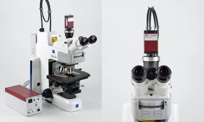
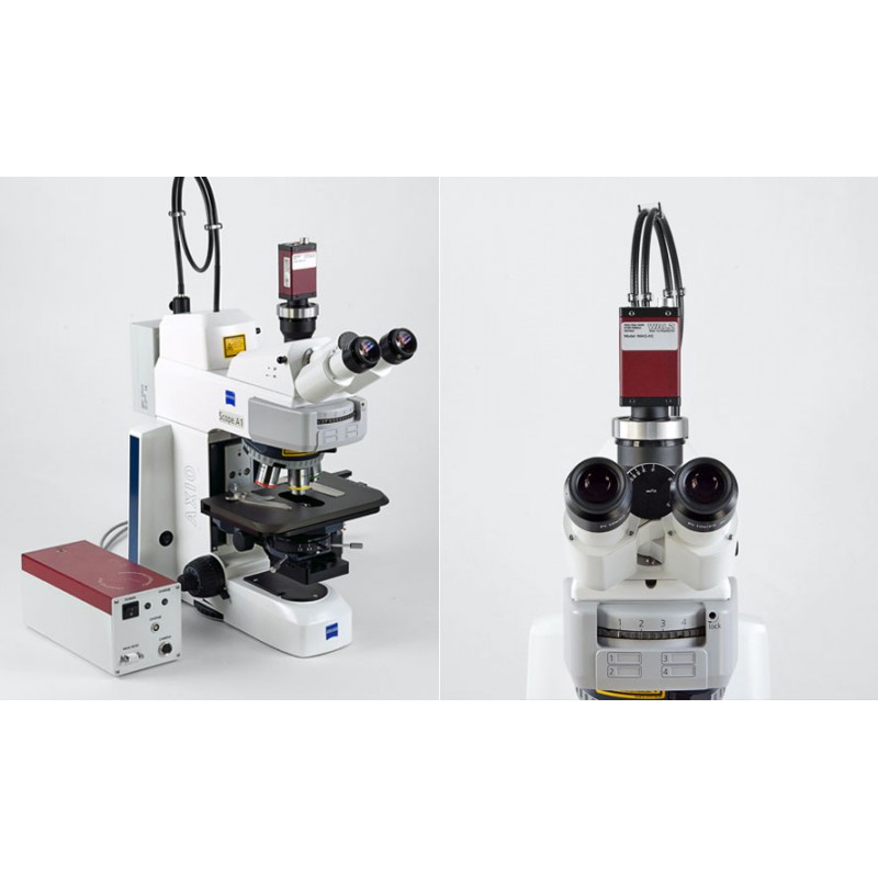
Microscope
• IMAG-AXIOSCOPE Axio Scope. A1 from Zeiss adapted for IMAGING-PAM applications. Detector filter RG665, dichroic mirror 420-640 nm, video adapter 60N-C 2/3' 0,5x and standard lens Zeiss Fluar 20x.
Computer
• IMAG-PC a brand-name notebook PC
Optional
• IMAG-AX-REF Reflector Module with Filter Set consisting of a beam splitter filter (420-640 nm) and a detector filter (665 nm), mounted in a Zeiss reflector module frame for convenient measurements using more than one LED Lamp Module.
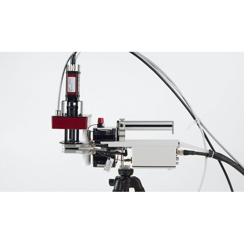
Due to its compact design, the MINI version is well suited for field applications. As the imaged area is much smaller than that of the MAXI version (17 x magnification of the area at the same resolution), maximal light intensities are higher, whereas power consumption is lower. For this reason the MINI version can easily be used as handy version independently from mains power.
The measuring head can also be mounted on the Standard Measuring Head 3010-S of the Portable Gas Exchange Fluorescence System GFS-3000 so that ImagingWin can directly communicate with GFS-Win for control and the exchange of data.
• IMAG-CG Multi Control Unit to connect Measuring Head and GigE –Vision® CCD camera as M-Series IMAGING-PAM MAXI, MINI and MICROSCOPY version including ImagingWin GigE Software.
• IMAG-MIN/B (blue measuring light version, 460 nm, standard applications)
• IMAG-MIN/R (red measuring light version, 620 nm, e.g. for cyanobacteria)
• IMAG-MIN/GFP (blue measuring light version, 470 nm, GFP imaging and PSII fluorescence) using a high sensitivity camera IMAG-K6, K6-MIN prime lens and Detector Filter Slide K6-MIN/FS
• IMAG-K7 (1/2" chip, 640 x 480 pixel) with K7-MIN prime lens (F1.4/f=16 mm)
• IMAG-K6 increased sensitivity (2/3" chip, primary resolution 1392 x 1040 pixels) with K6-MIN prime lens objective (F1.4/f=12.5 mm)
• all cameras need a mounting set K7-MIN/M or K6-MIN/M for robust coupling to the MINI-Head.
• IMAG-MIN/BK Leaf holder fixes a leaf to the MINI-Head sample frame
• ST-1010 compact tripod with UNC1/4-20 screw threads tripod head
• IMAG-MIN/ST fine drive with 120 mm traverse path for adjustment of the working distance coupled with a tripod adapter for mounting the MINI-Head onto a UNC1/4-20 screw threads tripod head
• IMAG-S Fine Drive Laboratory Stand with 50 mm traverse path high performance rack-and-pinion drive and 25 x 17 cm platform base.
• IMAG-PC a brand-name notebook PC
• IMAG-MIN/GFS Adapter for mounting MINI-Heads on Standard Measuring Head 3010-S of GFS-3000
Multi Control Unit
• IMAG-CG Multi Control Unit to connect Measuring Head and GigE–Vision® CCD camera as M-Series IMAGING-PAM MAXI, MINI and MICROSCOPY version including ImagingWin GigE Software.
Measuring Head
• IMAG-L470M Microscope LED Lamp Module 470 nm (blue) for fluorescence excitation of chlorophyll fluorescence
• IMAG-L625M Microscope LED Lamp Module 625 nm (red-orange) for fluorescence excitation of chlorophyll fluorescence of most algae groups including cyanobacteria
• IMAG-RGB Red-Green-Blue Microscopy LED Lamp Module allowing computer-assisted deconvolution of major algae groups. Fluorescence excitation and actinic illumination using red (620 nm), green (520 nm), blue (460 nm) or white light (mixed 620, 520 and 460 nm).
CCD Camera
• IMAG-K6 increased sensitivity (2/3' chip, primary resolution 1392 x 1040 pixels)
Microscope
• IMAG-AXIOSCOPE Axio Scope. A1 from Zeiss adapted for IMAGING-PAM applications. Detector filter RG665, dichroic mirror 420-640 nm, video adapter 60N-C 2/3' 0,5x and standard lens Zeiss Fluar 20x.
Computer
• IMAG-PC a brand-name notebook PC
Optional
• IMAG-AX-REF Reflector Module with Filter Set consisting of a beam splitter filter (420-640 nm) and a detector filter (665 nm), mounted in a Zeiss reflector module frame for convenient measurements using more than one LED Lamp Module.
.jpg)
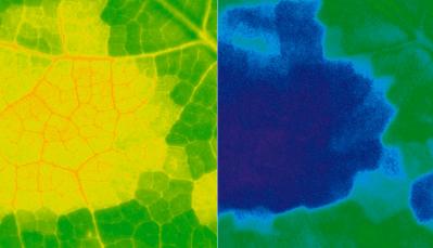
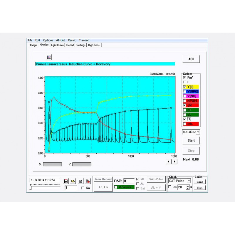
The IMAGING-PAM M-Series GigE instruments are fully controlled by the ImagingWinGigE software. When started, the ImagingWinGigE software opens with the image window that occupies most of the user surface showing the Ft value as starting parameter.
Values are represented in a false color scale ranging from black (0.0) to white (1.0) with red, orange yellow, blue and violet to purple in between. At first a standard AOI (area of interest) is already present after the start of the software. Different shapes and up to 100 AOIs can be defined. AOIs` positions can be moved by the new Edit function.
The kinetics window shows various parameter values for some or all AOIs of the currently chosen experiment plotted versus time. It serves for the evaluation of dynamic dark / light phenomena (Kautsky curve or Induction curve).
The customer can chose between 18 different parameters (Ft, Fo, Fm, F, Fm’, Fv/Fm, Y(II), Y(NPQ), Y(NO), PS/50, Abs, Red, NIR, NPQ/4, qN, qP, qL, Inh.) that could be displayed in the image window in different color modes. In this tab the alteration of the parameters can be observed in real-time during the experiment.
Some of the possible experiments are already preset in this and the following light curve window so that also the beginner finds an easy starting point for his first experiments. For advanced users it is also possible to program script files with more complex structure.
Some new features can be provided solely for the ImagingWinGigE software, not for the ImagingWin software suitable for FireWire camera versions of the IMAGING-PAM M-Series.
Easy light calibration using the ULM-500 Light Meter & Logger. ImagingWinGigE in communication with the ULM-500 provides an automated light calibration routine to generate a calibrated internal light list and furthermore offers to follow an external illumination.


![MC-100 Chlorophyll Concentration Meter [µmol m-2] with internal GPS](https://alphaomega-electronics.com/18349-home_default/mc-100-chlorophyll-concentration-meter-mol-m-2-with-internal-gps.jpg)
![MC-100 Chlorophyll Concentration Meter [µmol m-2] with internal GPS 2](https://alphaomega-electronics.com/1389-home_default/mc-100-chlorophyll-concentration-meter-mol-m-2-with-internal-gps.jpg)


